Standard anaesthesia practice involves the use of neuromuscular blocking agents (NMBAs) to facilitate intubation as well as improve surgical conditions by enabling muscle relaxation during the surgical procedure. Whenever NMBAs are given during anaesthesia, it is mandatory to monitor the degree of neuromuscular blockade (NMB).1 This can be done using subjective or objective methods of assessment. Subjective assessment, such as the evaluation of muscle strength or respiratory function, has proven inadequate as it lacks sensitivity and specificity. Furthermore, it is subject to interobserver variability. Therefore, clinical practice guidelines generally recommend objective assessment of neuromuscular transmission (NMT), using quantitative methods to measure TOF ratios.1
Why Quantitative Monitoring Makes Sense for Clinicians, Patients, and Budgets
Quantitative neuromuscular monitoring has benefitted clinicians, patients, and healthcare facilities alike:
-
Quantitative monitoring guides safe patient extubation
The incidence of residual neuromuscular block can be as high as 45%, even if a single dose of NMBA is used
The biggest challenge with the use of NMBAs is that their effect persists into the postoperative period. The incidence of residual neuromuscular block can be as high as 45%, even if a single dose of NMBA is used.2 Residual neuromuscular blockade in turn increases the risk of serious postoperative complications following extubation. These include aspiration, pneumonia, pharyngeal dysfunction, hypoxaemia, and airway obstruction. Such complications increase the length of stay of patients in the post-anesthesia care unit (PACU) and escalate care delivery costs.
Clinical guidelines recommend that for safe extubation, a minimum TOF ratio of 0.9 must be achieved.3 Quantitative monitoring grounds decision-making in a more sensitive and reliable evaluation of the TOF ratio. The result is markedly improved patient outcomes. In a randomised trial, Gatke et al. found that anaesthetists tended to extubate patients at least 2.5 minutes later when quantitative monitoring systems were used.4 This helped to decrease the incidence of residual muscle paralysis by 13.7%. In another randomised trial, Murphy et al. showed that compared to qualitative monitoring, the use of quantitative methods helped reduce cases of residual blockade by 4.5% and adverse respiratory events by 21%.5
-
It provides crucial data for appropriate intraoperative patient management
During surgical procedures, depth of neuromuscular blockade can be assessed using quantitative devices. The data guides the effective administration of repeat doses of NMBAs, preventing ‘bucking’ of patients during the procedure. Using optimal doses of NMBAs can help save time towards the end of a surgical procedure as well as avoid the need for reversal. All these factors contribute to freeing up operating room (OR) and PACU resources and cutting costs.
-
Quantitative monitoring enables optimal utilisation of reversal agents
Traditionally, acetylcholinesterase inhibitors such as neostigmine have been used to reverse NMB. However, the efficacy of these drugs in individuals is often unpredictable. One observational study documented that residual NMB occurred in at least 46% of patients in whom neostigmine was used.6 Moreover, such drugs have autonomic side effects, including increased respiratory secretions, bronchospasm, and bradycardia. The new reversal agent sugammadex gives more predictable results and does not have autonomic side effects. However, its high cost has been a hurdle to its more liberal use. When quantitative NMT monitoring is used, the depth of neuromuscular blockade can be assessed prior to reversal, and sugammadex can be reserved for deeper levels of blockade. Quantitative monitoring can thus help in optimising the use of reversal agents.
The Current Clinical Choices in Quantitative NMT Monitoring: AMG and EMG
Several methods have been described to quantitatively measure neuromuscular function. Mechanomyography (MMG), one of the earliest described techniques, directly measures the isometric force of muscle contraction. Output from MMG is precise and reproducible. However, it was primarily developed for research and is too cumbersome for clinical applications. It still remains the ‘gold standard’ for quantitative monitoring against which all the other methods and devices are compared.7 In medical practice, the two technologies that have found widespread adoption are acceleromyography (AMG) and electromyography (EMG).
How They Work—The Science of Monitoring
-
AMG
Acceleromyography is based on Newton’s second law of motion, which states that force is the product of mass and acceleration. Acceleration is detected by a piezoelectric sensor. The sensor, which has a constant mass, is attached to the muscle. When the muscle contracts, an acceleration voltage signal is detected by the sensor. The device uses this information to calculate the force of contraction and displays it on the monitor.
-
EMG
When a muscle contracts, it generates an electric signal called the compound action potential. This signal is directly proportional to the force of muscle contraction. An EMG device senses this electric signal. It does this through sensing electrodes that are placed on the surface of the muscle. The device estimates the force of contraction based on the strength of the electric signal and displays it on the monitor.
AMG: Advantages and Challenges
Acceleromyography is the most widely used technology for quantitative neuromuscular monitoring. One of the main reasons behind its wide clinical acceptance is its cost-effectiveness.  It is the least expensive of all the quantitative monitoring technologies available, owing to the fact that the piezoelectric sensor can be reused for multiple patients. Devices developed by several different manufacturers are available on the market. In addition, AMG technology has been extensively investigated in clinical studies. Therefore, its use is based on a sound body of clinical evidence.
AMG devices are available as portable, standalone units. They can be used at several sites, such as the thumb muscle (adductor pollicis), the foot muscle (flexor hallucis brevis), and eye muscles (orbicularis oculi and corrugator supercilii).
Acceleromyography is the most widely used and cost-effective technology for quantitative neuromuscular monitoring
AMG devices come with either one- or three-dimensional sensor technology. Devices with one-dimensional technology measure acceleration in a single dimension, while 3D devices measure acceleration in three dimensions. A big drawback of the 1D devices is the meticulous preparatory setup that they require. They need to be calibrated before each use. This is done by detecting the supramaximal current and correspondingly adjusting the twitch response to 100% to determine the full-scale deviation – a process that can take several minutes.
Fortunately, the 3D devices—such as the Stimpod NMS450X—do not suffer from this drawback and require no calibration. Supramaximal current can, and should still be determined – however this is now an automated process that takes less than 20 seconds.
Although some studies have suggested that the precision of AMG devices is increased when a preload is applied by returning the thumb to its original position after each stimulation, the evidence for this remains weak.8 A preload requires fitting a special hand adaptor. When the adductor pollicis is used, the patient’s hand and arm must be secured to the arm board of the operating table in such a way that thumb movements occur in a horizontal direction.
AMG devices also exhibit the “reverse fade” effect, a similar effect that is also noticeable to a lesser extent with mechanomyography (MMG) devices without a preload. When 1.0 is the ideal baseline value, values of 1.10 to 1.47 have been recorded in literature.9 A high baseline value would naturally affect the TOF ratio measured during recovery. Some experts have suggested that instead of 0.9, a TOF ratio of 1.0 or higher should be obtained prior to extubation. Another method suggested to overcome this effect is to normalise the TOF values obtained against the baseline. This methodology however can result in a high time cost at the start of the procedure waiting for a stable baseline, rendering this option impractical in most cases.
EMG: Advantages and Challenges
 Electromyography is the most ‘physiological’ way of assessing muscle function and is, therefore, more precise than AMG. Since the electric signal is measured rather than the force of muscle contraction, free movement of the muscle is not required. Hence, there is no need for a special ‘setup’ as with the AMG. EMG devices can be used when rigid patient positioning is required. For example, when the arms need to be ‘tucked’ close to the body or during robotic surgery. The increasing adoption of robotic surgery creates a special use case for EMG devices.
Unlike AMG, EMG does not have the ‘reverse fade’ effect. A recent study compared EMG and AMG monitors against the gold standard MMG. The authors found that an AMG monitor would frequently produce TOF ratios > 1.0, yet neither EMG nor MMG (with a preload) exhibited this phenomenon to any significant extent.10
EMG devices can be used when rigid patient positioning is required
A disadvantage of EMG devices is the possibility of interference from other electrical devices, such as electrocautery. The use of such equipment may distort the electric signal and give incorrect readings. Output may also be affected by temperature changes, as reduced temperatures tend to amplify EMG responses. In contrast, the Stimpod NMS450X instantaneously detects interference from electrocautery, automatically pauses the EMG measurement and resumes when the interference is gone.
Furthermore, EMG devices depend on special electrodes, which are often expensive and difficult to source. This amplifies per case costs, which is a major limiting factor to the widespread use of EMG in PACUs and ICUs. It is also the reason why there is not widespread adoption of commercial, portable devices that use EMG technology exclusively.
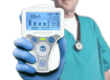
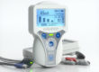
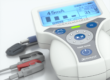
Stimpod NMS450X: The only standalone monitor with both AMG and EMG options at Point of Care
The Stimpod NMS450X combines both AMG and EMG capabilities in a standalone, portable device. It is truly disruptive technology as a comparable device simply doesn’t exist. It brings NMT monitoring science from the lab to point of care.
Choosing between AMG and EMG can be baffling—unless you have the Stimpod NMS450X, which offers the best of both:

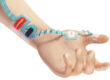
Stimpod’s unique EMG electrode design is unmatched in the industry:
- Patented ripple design to improve ambidextrous and varying patient placement as well as tension transfer
- Specialty ‘skin-like’ material for maximum hold during long surgeries and patient movement
- Thin, ultra-flexible design for quick, effective placement for any setup
- Superior signal processing for data precision
As a whole, it is the complete quantitative NMT monitoring solution:
- Does not need calibration: It is quick and easy to set up, and no normalisation is needed before use.
- Cost-effective sensors: Hospitals now have the option of choosing their cost of care between re-usable AMG and single-use EMG at the point of care.
- Versatile: The Stimpod is mountable for complete OR intraoperative use or in-hand portable for bedside use in the PACU or ICU.
- A complete neuromuscular monitoring solution: Complete monitoring for any patient in any setting. 100% hospital settings, 100% provider preference, 100% surgery types in combination with full case, automatic monitoring.
In summary, the Stimpod NMS450X enables you to leverage the power of both AMG and EMG for reliable and accurate quantitative NMT monitoring at your facility. What this translates into is better health outcomes for your patients and a substantial reduction in costs for you. If you would like to know more, please get in touch and we can discuss your specific needs.
References
- Checketts MR, Alladi R, Ferguson K, et al. Recommendations for standards of monitoring during anaesthesia and recovery 2015: Association of Anaesthetists of Great Britain and Ireland. Anaesthesia. 2016;71(1):85-93. doi:10.1111/anae.13316
- Debaene B, Plaud B, Dilly MP, Donati F. Residual Paralysis in the PACU After a Single Intubating Dose of Nondepolarizing Muscle Relaxant with an Intermediate Duration of Action. Anesthesiology 2003;98: 1042–8.
- Popat M, Mitchell V, Dravid R, Patel A, Swampillai C, Higgs A. Difficult Airway Society Guidelines for the management of tracheal extubation. Anaesthesia 2012; 67: 318– 40.
- Gätke MR, Viby-Mogensen J, Rosenstock C, Jensen FS, Skovgaard LT. Postoperative muscle paralysis after rocuronium: less residual block when acceleromyography is used. Acta Anaesthesiol Scand. 2002;46(2):207-213. doi:10.1034/j.1399-6576.2002.460216.x
- Murphy GS, Szokol JW, Marymont JH, et al. Intraoperative acceleromyographic monitoring reduces the risk of residual neuromuscular blockade and adverse respiratory events in the postanesthesia care unit. Anesthesiology. 2008;109(3):389-398. doi:10.1097/ALN.0b013e318182af3b
- Aytac I, Postaci A, Aytac B, et al. Survey of postoperative residual curarization, acute respiratory events and approach of anesthesiologists. Braz J Anesthesiol. 2016;66(1):55-62. doi:10.1016/j.bjane.2012.06.011
- Murphy GS. Neuromuscular Monitoring in the Perioperative Period. Anesth Analg. 2018;126(2):464-468. doi:10.1213/ANE.0000000000002387
- Claudius C, Viby-Mogensen J, Warner DS, Warner MA. Acceleromyography for Use in Scientific and Clinical Practice: A Systematic Review of the Evidence. Anesthesiology 2008; 108:1117–1140 doi: https://doi.org/10.1097/ALN.0b013e318173f62f
- Suzuki T, Fukano N, Kitajima O, Saeki S, Ogawa S: Normalization of acceleromyographic train-of-four ratio by baseline value for detecting residual neuromuscular block. Br J Anaesth 2006; 96:44–7.
- Bowdle A, Bussey L, Michaelsen K, et al. A comparison of a prototype electromyograph vs. a mechanomyograph and an acceleromyograph for assessment of neuromuscular blockade. Anaesthesia. 2020;75(2):187-195. doi:10.1111/anae.14872
Contributors
Roche Janse van Rensburg, Maruschka van der Bank, Lourie Höll
Enquiries
Maruschka van der Bank
Product Specialist
maruschka@xavant.com